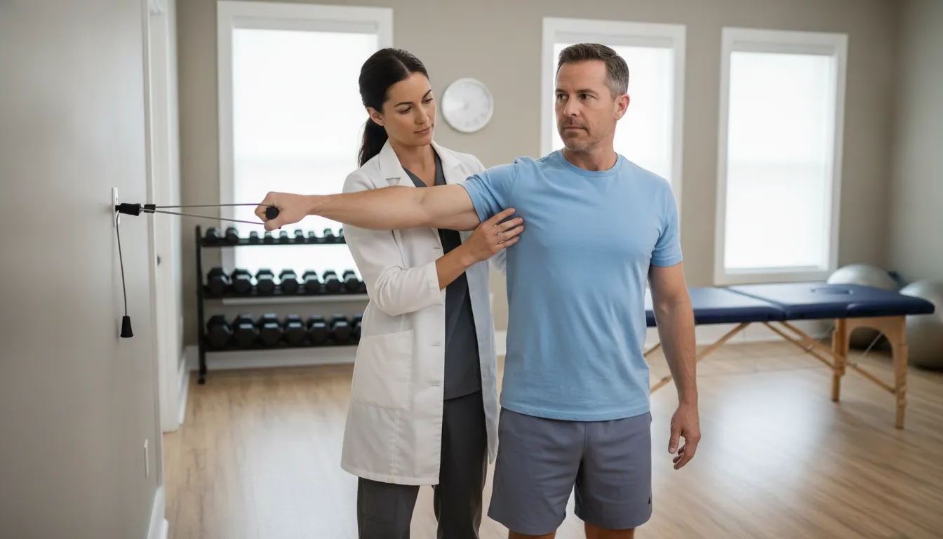
Foot Drop Treatment and Rehabilitation: Exercises That Help
Foot drop often starts with a subtle sign. You may scuff one shoe, catch your toes on a rug, or feel more tired after walking. Over time, these changes can affect your balance and confidence. Because foot drop may stem from a compressed nerve, spinal stenosis, neuropathy, stroke, injury, or another condition, home exercises alone may not be enough. A timely evaluation helps your care team identify the source and determine the safest next step. Physical medicine and rehabilitation for foot drop may support recovery through targeted exercises, gait retraining, balance practice, bracing, and strategies that reduce fall risk while you work toward better mobility.
Key Takeaways
- Treat new foot weakness as a medical concern: Toe dragging, foot slapping, numbness, or a high-stepping gait may point to a nerve, spine, muscle, or neurological problem that requires evaluation.
- Build rehabilitation around the underlying cause: Rehabilitation may combine ankle and leg strengthening, flexibility work, gait practice, balance training, bracing, and assistive devices to support safer movement and independence.
- Know when to seek immediate care: Sudden weakness, facial drooping, speech changes, severe back pain, saddle numbness, bowel or bladder changes, worsening symptoms, or repeated falls require prompt medical attention.
What Is Foot Drop and What Causes It?
Foot drop is difficulty lifting the front of the foot while walking. It describes a movement problem, not a diagnosis. The weakness may begin in the muscles, peripheral nerves, lower back, spinal cord, or brain. Identifying the source is essential because treatment may include rehabilitation, an ankle-foot orthosis, nerve or spine care, medication changes, or another medical intervention.
Some people notice foot drop after an injury, while others develop it gradually. Toe catching, foot slapping, and a new need to lift the knee higher are signs worth evaluating, especially if they interfere with exercise, stairs, or daily activities. Do not assume new walking difficulty is simply part of aging. A timely assessment can identify treatable causes and help reduce fall risk.
Foot drop signals weakness, not a diagnosis
Foot drop occurs when the front of the foot does not lift normally. You may notice your toes dragging across the floor, your foot slapping down, or your knee rising higher than usual to help the foot clear the ground. Symptoms can affect one leg or, less often, both legs.
The term describes how the foot moves, but it does not identify the source of the problem. Compression of the peroneal nerve near the knee, a pinched nerve in the lower back, peripheral neuropathy, or a neurological condition can create similar walking changes. A clinician will review your medical history and assess strength, sensation, reflexes, and gait. The Mayo Clinic’s guide to foot drop explains why finding the underlying cause helps determine appropriate care.
How dorsiflexion and toe extension work
Dorsiflexion brings the top of your foot and toes closer to your shin. This movement helps your toes clear the floor as your leg swings forward. Toe extension, or lifting the toes, also supports foot clearance and helps position the foot before it contacts the ground.
Muscles in the front and outer part of the lower leg work together to create these movements. Nerves carry instructions from the brain and spinal cord to those muscles. If the muscles are weak, nerve signals are disrupted, or the ankle is stiff, the foot may not lift high enough during walking. The Mayo Clinic describes the role of dorsiflexion in walking, which can help you understand why even a small loss of movement affects balance and mobility.
Common causes: nerve compression, radiculopathy, stenosis, neuropathy, stroke, and injury
The peroneal nerve, also called the common fibular nerve, controls several muscles that lift and turn the foot outward. Pressure or injury near the outside of the knee can affect this nerve. Prolonged pressure, trauma, surgery, or frequent leg crossing may contribute in some cases.
Foot drop may also result from radiculopathy, in which a nerve root in the lower back becomes irritated or compressed. Lumbar spinal stenosis can narrow the spaces around spinal nerves and contribute to weakness, numbness, pain, or walking changes. Peripheral neuropathy, stroke, multiple sclerosis, and injuries affecting the leg, ankle, or lower back are other possible causes. The National Institute of Neurological Disorders and Stroke explains how stroke can affect movement and strength.
How sudden and gradual onset differ
Sudden foot drop requires prompt medical attention, especially when it appears with facial drooping, speech difficulty, confusion, severe headache, or weakness in an arm. These symptoms may signal a stroke and warrant emergency care. Sudden weakness can also follow an injury or occur when a nerve or spinal condition changes quickly.
Gradual foot drop may develop over weeks or months. Early signs can include occasional toe catching, reduced walking endurance, numbness, or difficulty on uneven ground. This pattern may occur with neuropathy, spinal stenosis, or a slowly progressing nerve problem. Even gradual symptoms deserve evaluation. Orthopedic Reviews discusses sudden and gradual foot drop presentations, including how the pattern of onset can help guide further assessment.
Why the cause guides treatment
Rehabilitation can address strength, joint mobility, balance, walking mechanics, and confidence, but the plan should reflect the reason foot drop developed. For example, care for nerve compression may focus on reducing pressure and protecting the nerve. Rehabilitation for spinal stenosis may emphasize posture, walking tolerance, and lower-body strength. With neuropathy, treatment may place greater attention on sensation, skin checks, footwear, and fall prevention.
Recovery also depends on whether the affected nerve or muscle can heal. Some people regain useful strength as the underlying condition improves. Others may need a brace, functional electrical stimulation, specialist treatment, or long-term adaptations. The Mayo Clinic notes that foot drop may improve when its cause is treated successfully. A thorough evaluation can establish practical goals and clarify which concerns rehabilitation can address directly.
What Symptoms and Fall Risks Does Foot Drop Cause?
Foot drop affects your ability to lift the front of your foot while walking. This movement, called dorsiflexion, helps your toes clear the ground during each step. When the muscles or nerves involved in dorsiflexion are weak, your foot may drag, slap the floor, or catch on small obstacles. Foot drop is a sign of an underlying condition, not a diagnosis by itself. The cause may involve a nerve, the lower back, the muscles, or the brain, so symptoms can differ from one person to another.
Fall risk depends on more than ankle weakness. Reduced sensation, poor balance, limited joint movement, vision problems, and fatigue can all make walking less secure. A flat floor at home may feel manageable, while stairs, curbs, dim lighting, and uneven ground may expose problems with foot clearance. Recognizing these changes early can help you seek an evaluation and make practical safety adjustments before foot drop limits your daily activities.
Toe catching, foot slapping, and high-stepping
A common sign of foot drop is catching your toes on the floor while walking. You may notice scuffed shoes, stumble over rugs, or concentrate more than usual on lifting your foot. Some people develop a foot slap, where the front of the foot drops suddenly instead of landing in a controlled way. This may create a louder sound with each step and indicate reduced ankle control.
When the foot does not clear the ground, your body may lift the knee higher to avoid dragging the toes. This creates a high-stepping pattern, often called a steppage gait. The Orthopedic Reviews overview of foot drop explains how this compensation can develop when dorsiflexion is weak. Frequent toe catching or a new foot slap deserves medical attention, particularly if the change appeared suddenly or is getting worse.
Numbness, tingling, pain, and reduced foot awareness
Foot drop may occur with numbness, pins-and-needles sensations, burning, or pain in the foot and lower leg. These symptoms can result from irritation or damage along a nerve pathway, including a nerve near the knee or a nerve root in the lower back. Some people have weakness without pain, while others notice back, hip, leg, or foot discomfort at the same time.
Reduced sensation can make walking less secure because you may not clearly feel where your foot lands. You may miss a small change in the floor, pressure from a shoe, or irritation from a brace. Ortho Foot & Ankle’s guide to drop foot lists difficulty lifting the front of the foot, dragging during walking, numbness or tingling, and lower-leg weakness among common signs. Check your feet regularly when sensation is reduced, especially if you have neuropathy or diabetes.
Difficulty with stairs, curbs, uneven ground, and transfers
Foot drop often becomes more noticeable when the walking surface changes. Stairs require sufficient foot clearance and ankle control to place the foot securely on each step. Curbs, gravel, grass, and cracked pavement create similar challenges. If your toe catches, you may lose balance before you have time to reach for a railing or adjust your step.
Transfers can also feel less steady. Standing from a chair, getting out of a car, and turning in a narrow space require coordination between your feet, legs, and trunk. Weakness or reduced sensation may cause you to place the affected foot awkwardly or shift too much weight to the stronger side. Advanced Reconstruction describes the effects of foot drop on mobility as symptoms progress. Use handrails, slow down, and keep walkways clear while arranging an evaluation.
Compensatory hip and knee movements
When the ankle cannot lift the foot normally, other joints may work harder to keep the toes from catching. You may raise your hip, bend your knee more, swing the leg outward, or lean your trunk while walking. These adjustments can help you move forward, but they may also make your gait less efficient and place extra demand on the hip, knee, pelvis, or lower back.
Over time, these patterns may contribute to fatigue, soreness, or the feeling that walking takes more effort than it should. Compensation can also make it harder to stay balanced when turning or changing speed. A clinician can assess how your entire body moves, rather than focusing only on the ankle. Treatment may need to address hip and core strength, balance, flexibility, and walking mechanics along with the muscles that lift the foot.
How foot drop affects exercise, confidence, independence, and daily function
Foot drop can change which activities you feel comfortable doing. You may avoid walks, trails, exercise classes, yardwork, or travel because you are concerned about tripping. Reduced activity can then contribute to more weakness, stiffness, and loss of conditioning. Routine tasks, including carrying groceries, showering, or moving through a busy home, may require extra attention.
The emotional impact matters, too. A near-fall can make you cautious, while repeated stumbles may reduce your confidence in your ability to remain independent. Rehabilitation can focus on practical goals, such as walking to the mailbox, using stairs, returning to strength training, or getting up from the floor. A comprehensive foot drop rehabilitation case report describes improvements in walking pattern, balance, lower-limb strength, and range of motion after targeted care. Results depend on the cause and severity, but an individualized plan may help you move more safely and maintain daily function.
How Do Clinicians Evaluate Foot Drop Before Rehabilitation?
Before rehabilitation begins, clinicians need to understand why foot drop developed and how it affects your movement. Foot drop is a sign of weakness that may involve the brain, spinal cord, nerve roots, peripheral nerves, muscles, or joints. Because the cause can vary, exercises alone may not address the problem.
A thorough evaluation helps your care team identify fall risks, choose appropriate exercises, and determine whether you need additional medical treatment. It may include a physical examination, strength and movement testing, gait analysis, and diagnostic studies. Your provider may also coordinate care with neurology, spine, pain, or other specialists.
The evaluation should account for your daily life, not just muscle grades or test results. Your ability to walk through your home, climb stairs, exercise, work, travel, and remain independent all help shape the treatment plan.
Review your medical history and neurological status
Your clinician will ask when the weakness began, whether it appeared suddenly or gradually, and whether it has changed over time. Be prepared to discuss back or leg pain, numbness, tingling, recent injuries, surgeries, illnesses, medication changes, and any history of diabetes, stroke, neuropathy, or spine conditions.
Your provider may also ask about falls, near-falls, changes in exercise tolerance, and activities that have become difficult. During the neurological examination, they may compare both legs, observe muscle control, and look for patterns that suggest nerve root or peripheral nerve involvement. The Mayo Clinic’s overview of foot drop diagnosis explains that clinicians commonly watch patients walk and check for weakness in the leg muscles.
Test foot, ankle, knee, hip, and core strength
Ankle weakness is central to foot drop, but it is only one part of the assessment. Your clinician may test dorsiflexion, which lifts the front of the foot, along with toe extension, eversion, and plantarflexion. They may also assess your knee, hip, and core because weakness higher in the leg can affect balance and walking mechanics.
Strength testing often involves moving against gravity and gentle resistance. A severely weak ankle may show only a small muscle contraction, while moderate weakness may allow movement but not against resistance. Clinicians may record each muscle group using a standard grading system so they can compare your results over time. This published case report on foot drop rehabilitation demonstrates how ankle strength measurements can guide rehabilitation and help track progress.
Check sensation, reflexes, mobility, and muscle tone
Your care team may use light touch, pressure, or other simple tests to check sensation in your foot and leg. Numbness or reduced foot awareness can make it harder to feel the ground, increasing the risk of tripping on stairs, curbs, or uneven surfaces. Sensory changes may also provide clues about the affected nerve or nerve root.
Clinicians may check reflexes at the ankle and knee, evaluate muscle tone, and measure how far your ankle can move. They will look for stiffness, contractures, swelling, pain, or a fixed foot position that interferes with clearance. An absent ankle reflex or an inability to actively lift the foot can be important findings, as shown in this clinical foot drop rehabilitation case. These results help determine which exercises and mobility supports are appropriate.
Assess gait, balance, foot clearance, and fall risk
Your clinician may watch you walk at different speeds, turn, step over an object, and move across various surfaces. They will look for toe catching, foot slapping, high-stepping, shortened steps, uneven weight shifting, and extra movement at the hip or knee. These patterns show how your body is compensating for limited foot control.
Balance testing may include standing with your feet together, shifting weight, rising from a chair, or walking while turning your head. Your provider may also ask about falls, near-falls, confidence, and your ability to manage stairs and curbs. Foot drop can change your walking pattern early on, so recognizing these changes can help prevent injuries. This overview of foot drop warning signs describes how altered walking may affect safety and quality of life.
Use EMG, nerve studies, imaging, or blood tests when needed
The physical examination may identify foot drop, but additional testing can help locate its cause. Electromyography and nerve conduction studies assess how nerves and muscles respond. These tests may help distinguish a compressed nerve root from a peripheral nerve problem or another neurological condition.
Imaging may include an MRI or another scan of the spine, brain, knee, or lower leg, depending on your symptoms. Blood tests may be useful when clinicians suspect diabetes, nutritional deficiency, inflammation, infection, or another systemic condition. Testing should match your medical history and examination rather than follow the same process for everyone. If your foot is held in an abnormal position, your clinician may also determine whether the deformity is flexible or rigid, an issue discussed in this review of foot drop treatment.
Refer you to neurology, spine, pain, or other specialists
Some people can begin rehabilitation after an initial evaluation. Others may need coordinated medical care before or during rehabilitation. A neurologist can assess nerve and muscle disorders. A spine specialist can evaluate stenosis, a disc problem, or nerve root compression. A pain physician can help manage symptoms that make walking or exercise difficult.
Referral may be appropriate when weakness is progressing, symptoms affect both legs, pain is severe, or the examination suggests a condition outside the clinician’s scope. In some cases, inflammatory or blood vessel disorders can contribute to foot drop, so a broader assessment is necessary. Your care team may work together to determine whether rehabilitation, bracing, medication, injections, nerve treatment, or surgery should be part of your care.
Set goals for walking, exercise, recovery, and independence
The evaluation should lead to a practical plan, not just a list of test results. Your goals might include walking without catching your toes, climbing stairs, returning to golf or hiking, standing from a chair, or exercising with less fear of falling. If you use a brace or cane, your plan may also focus on comfort, confidence, and safe device use.
Your clinician may record strength, walking speed, balance, range of motion, and the number of trips or falls so progress is easier to measure. A case report on rehabilitation after lumbar spine treatment describes how a tailored rehabilitation program can support walking and recovery in a person with foot drop. Goals may change as your symptoms, abilities, and daily needs change.
How Can Rehabilitation Help Foot Drop?
Rehabilitation for foot drop focuses on safer movement, better leg control, and maintaining independence. The right plan depends on the cause of weakness, how long symptoms have been present, how much movement you have, and what you want to do. Your goals may include walking without tripping, climbing stairs, returning to exercise, or moving around your home with more confidence.
A clinician may combine mobility exercises, strengthening, gait training, balance work, hands-on treatment, and recommendations for braces or assistive devices. Rehabilitation cannot repair every type of nerve damage, but it may improve joint motion, muscle control, walking mechanics, and day-to-day function. It should also work alongside medical care when foot drop may be related to nerve compression, a spine condition, stroke, injury, or another condition that requires additional evaluation.
Build a plan around the cause and your goals
Foot drop is a symptom rather than a single diagnosis. It may result from nerve compression near the knee, a pinched nerve in the lower back, peripheral neuropathy, stroke, injury, or a condition affecting the muscles or brain. Your clinician will consider your medical history, strength, sensation, reflexes, joint movement, and walking pattern when planning treatment.
Your personal goals shape the plan, too. One person may want to walk safely across an uneven lawn, while another may hope to return to hiking, cycling, or strength training. Treatment may focus on clearing the foot during each step, improving stair control, reducing pain, or increasing endurance. According to the Mayo Clinic’s guidance on foot drop treatment, improvement often depends on whether the underlying cause can be treated.
Restore ankle movement and reduce stiffness
When the ankle has little active movement, treatment may begin with passive or assisted exercises. These movements guide the foot through a comfortable range without asking weak muscles to do more than they can manage. They may help maintain flexibility, reduce stiffness, and lower the risk of a contracture, which occurs when a joint becomes fixed in a shortened position.
Your clinician may recommend gentle calf stretches, ankle range-of-motion exercises, and positioning strategies for rest or sleep. Hands-on techniques may also help address tight muscles or restricted ankle movement. The goal is not to force the foot into a stretch. Slow, controlled movement is generally more appropriate, especially when sensation is reduced. Learn how passive foot drop exercises work, but ask a clinician to demonstrate the safest options for you.
Strengthen the foot, ankle, lower leg, hip, knee, and core
Foot drop may involve more than the muscles that lift the front of the foot. Weakness in the calf, ankle stabilizers, hip, knee, or core can make it harder to control the leg while walking. Rehabilitation may start with small, supported movements and progress to resistance exercises, standing drills, and functional activities.
Your program may target dorsiflexion, which lifts the foot upward, and eversion, which turns the sole outward. It may also include bridges, hip abduction, sit-to-stands, and controlled step-ups. Stronger hips and a steadier trunk can improve leg alignment and reduce compensatory movements. A case report on comprehensive rehabilitation for foot drop describes a program that combined strengthening, stretching, balance training, gait work, and core exercises.
Retrain foot clearance and coordination
Strength alone may not restore a safe walking pattern. Your nervous system also needs repeated practice coordinating the ankle, knee, and hip at the right time. A clinician may use marching drills, toe lifts, stepping over low objects, treadmill practice, or slow walking with visual feedback to help you clear the ground.
You may begin in a supported position, such as sitting or standing near a stable surface. As your control improves, the clinician can add turns, stairs, uneven surfaces, or another simple task. Repeated movement practice may support neuroplasticity, the brain’s ability to relearn movement skills. Foot drop exercise guidance can help explain how consistent practice supports more coordinated movement.
Adapt rehabilitation for neuropathy, spine conditions, joint problems, pain, and fatigue
Two people with foot drop may need very different treatment. If neuropathy reduces sensation, care should include daily foot checks, balance strategies, and exercises that do not depend entirely on feeling the ground. If a lumbar spine condition affects a nerve root, your clinician may adjust positions and movements to avoid increasing back or leg symptoms.
Arthritis, hip weakness, knee pain, and limited stamina can also change how exercises are performed. Your clinician may use seated or supported activities, shorter sessions, rest periods, or water-based exercise when standing work is too demanding. Foot drop may involve the brain, muscles, peripheral nerves, or L4 and L5 nerve roots, so the broader symptom pattern matters when evaluating possible causes.
Clarify what rehabilitation can improve and what needs medical care
Rehabilitation may improve mobility, strength, balance, walking mechanics, and safety. It may also help you maintain joint motion while an affected nerve recovers. However, rehabilitation does not replace medical care when weakness is severe, rapidly worsening, or linked to significant nerve compression.
Ask your clinician what changes they expect and when additional evaluation may be appropriate. Severe foot drop, new neurological symptoms, or weakness that continues to progress may require assessment by a neurologist, spine specialist, or another physician. Testing or procedures may be necessary to address the cause. Clinical guidance on foot drop exercises notes that exercise may have limited effects when nerve damage or compression is severe.
Progress your home exercise program safely
A home program should match your current ability. If you have very little active ankle movement, begin with passive or assisted exercises rather than forcing resisted lifts. Your clinician can show you how many repetitions to complete, how often to practice, and which symptoms mean you should stop.
Progress may involve adding repetitions, increasing resistance, standing with less support, or practicing more challenging walking tasks. Change one part of the program at a time so you can tell how your body responds. Keep a stable surface nearby, wear secure shoes, and avoid exercises that increase your risk of tripping. Guidance on progressing foot drop exercises recommends starting with easier movements and advancing gradually, especially when weakness or balance problems are significant.
Which Foot Drop Exercises Improve Mobility and Strength?
The most useful foot drop exercises address more than the ankle. They can help you lift the front of your foot, control the lower leg, stabilize the hips and trunk, and respond to changes in the ground beneath you. Together, these skills support safer walking, improved balance, and greater confidence with everyday activities.
A clinician should choose exercises based on the cause and severity of your foot drop. Weakness related to a compressed nerve, lumbar spine condition, neuropathy, stroke, or injury may require a different plan. Your clinician can also determine whether you need an ankle-foot orthosis, electrical stimulation, or another form of support during exercise.
Perform each movement slowly and use a counter, sturdy chair, or wall for support as needed. Mild muscle fatigue may be expected, but sharp pain, dizziness, worsening numbness, or unsafe movement means you should stop and contact your medical team. If you have new or rapidly worsening weakness, seek medical evaluation before beginning a home exercise program.
Practice assisted and active ankle dorsiflexion and toe lifts
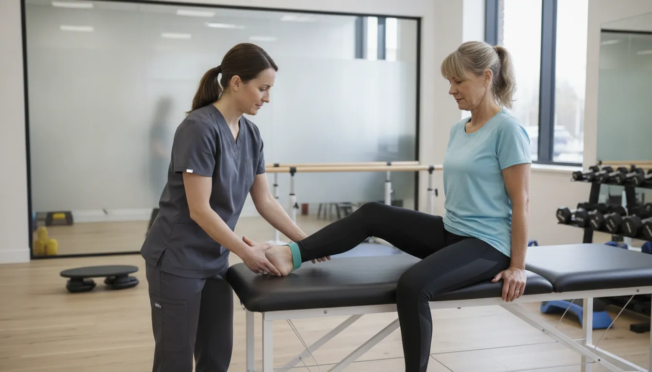
Ankle dorsiflexion is the movement of lifting your toes toward your shin. It helps clear the foot from the floor during walking and supports a more controlled heel strike. To practice, sit with your leg supported and slowly pull your toes upward. Hold briefly, then lower your foot with control.
If you cannot lift the foot independently, loop a towel or belt around the forefoot and use it to provide gentle assistance. Let your muscles do as much of the work as possible, then use the towel to complete the movement. You can also practice lifting the toes while keeping the heel on the floor. Flint Rehab’s foot drop exercise guide describes assisted and active drills that can be adapted to your ability.
Avoid forcing the ankle or jerking the foot upward. The goal is controlled practice, not simply moving through the largest range possible.
Stretch the calf and improve ankle range of motion
A tight calf can make it harder to bring your foot upward and may limit your ability to place the heel on the ground. Gentle calf stretching can help maintain ankle flexibility and reduce stiffness that interferes with walking. A clinician may recommend a wall stretch, seated towel stretch, or another position suited to your balance and sensation.
For a standing wall stretch, place the affected leg behind you with the heel down and toes facing forward. Keep the back knee straight for one version, then bend it slightly to target the deeper calf muscle. Lean forward until you feel a mild stretch, not pain.
Hold the position for about 20 to 30 seconds, if comfortable, and repeat as directed. Advanced Reconstruction’s foot drop exercise recommendations emphasize maintaining calf flexibility and ankle range of motion. Do not bounce or stretch aggressively, especially if you have reduced sensation.
Strengthen dorsiflexion, eversion, and the lower leg
Once you can move the foot with some control, strengthening exercises may help improve ankle stability. Dorsiflexion focuses on lifting the foot upward, while eversion involves turning the sole slightly outward. Eversion can engage the peroneal muscles, which help control the foot during walking and on uneven surfaces.
A resistance band may be useful, but only if your clinician confirms that you can perform the movement safely. Anchor the band securely and move through a comfortable range without rushing. You may also practice lifting the toes against light resistance or holding the foot in a neutral position.
Your clinician may begin with gravity-minimized exercises, manual assistance, or a small number of repetitions before adding resistance. Flint Rehab’s examples of ankle inversion and eversion exercises can be adjusted to your strength level. Stop if your foot begins to turn sharply, your symptoms increase, or you start compensating with your hip.
Train the hips, knees, and core for better leg control
Foot clearance depends on the entire leg, not only the ankle. Weak hip flexors, thigh muscles, or trunk muscles can make it harder to lift and place the leg safely. Exercises such as seated marching, straight-leg raises, bridges, and supported hip abduction may help develop the strength needed for more controlled steps.
Your clinician may also include knee extension exercises, mini-squats, or gentle core stability work. Stronger hips and knees can reduce the need to swing the leg outward or lift the hip excessively when walking. Better trunk control may help you remain steady when your foot does not respond quickly.
The right exercises depend on your balance, back symptoms, joint health, and sensation. A case report on comprehensive foot drop rehabilitation included hip, knee, and core strengthening to support improved leg control. Exercises should remain controlled and should not increase radiating pain or neurological symptoms.
Practice sit-to-stands, step-ups, and weight shifts
Functional exercises connect strength gains to the activities you perform every day. Sit-to-stands train the hips, knees, and trunk while helping you practice a controlled transition from sitting to standing. Use a firm chair, place both feet evenly on the floor, and push through your legs rather than pulling on the chair.
Step-ups can help prepare you for curbs and stairs, but begin with a low step, a secure railing, and supervision if needed. Focus on placing the whole foot securely before shifting your weight. If lifting the affected leg is difficult, your clinician may lower the step or choose a different exercise.
Weight-shifting drills are another option. While holding stable support, slowly move your weight from side to side and forward and backward. These movements help you practice loading the affected leg without taking a full step. Comprehensive foot drop rehabilitation research includes functional exercises such as sit-to-stands and step-ups.
Improve balance and proprioception with support
Proprioception is your awareness of where your foot and leg are positioned. Foot drop, numbness, and weakness can reduce this awareness, making uneven ground and quick changes in direction more difficult. Supported balance exercises give you a controlled way to practice responding to these challenges.
Begin with both feet on the floor while holding a counter or sturdy rail. Progress to placing one foot slightly ahead of the other, shifting weight onto the affected side, or briefly lifting one foot. Keep the support within reach, and have another person nearby if you are unsteady.
Balance work should challenge you without creating a fall risk. Do not begin with your eyes closed, unstable surfaces, or unsupported single-leg standing. Flint Rehab’s balance recommendations include supported single-leg practice to improve stability and foot awareness. Your clinician can determine when it is safe to make the exercise more difficult.
Match exercise intensity and progression to your ability
Foot drop exercises should match your current strength, sensation, balance, and energy level. Someone with mild weakness may tolerate resistance bands and standing drills, while someone with significant weakness may need seated exercises and hands-on assistance first. Starting at the appropriate level helps you practice quality movement without unnecessary strain.
Progression may involve adding repetitions, increasing hold times, reducing hand support, introducing resistance, or practicing on more challenging surfaces. Change only one part of the exercise at a time. If your foot starts dragging, your knee buckles, or you rely on large compensatory movements, the exercise may be too difficult.
Keep a simple record of your exercises, symptoms, walking tolerance, and near-falls. This gives your clinician useful information for adjusting your program. Advanced Reconstruction’s guidance on tailoring foot drop exercises recommends matching activity to your current ability and increasing intensity gradually. Consistent practice with safe form is more valuable than pushing through a demanding routine.
How Do Gait Training and Supportive Treatments Help?
Gait training helps you relearn safer, more efficient walking when foot drop affects foot clearance, balance, or leg control. A clinician observes how your foot, ankle, knee, hips, and trunk work together, then builds treatment around the movements that challenge you most. Sessions may include stepping over obstacles, walking on uneven surfaces, turning, climbing stairs, and rising from a chair.
Supportive treatments can make movement safer while strength and coordination improve. An ankle-foot orthosis, functional electrical stimulation, a cane, or a walker may reduce tripping and help you remain active. These tools do not treat every cause of foot drop, so they work best alongside a medical evaluation and personalized rehabilitation plan. The Mayo Clinic’s guidance on foot drop treatment explains why treatment depends on the underlying cause.
Retrain heel strike, foot clearance, cadence, and stair walking
Foot drop can change how your foot lands and make your toes catch the floor as your leg swings forward. Gait training breaks walking into smaller, repeatable skills. You may practice lifting your toes, bringing the affected leg forward, placing the heel down first, and shifting your weight with control.
Your clinician may also adjust your cadence, or step rate. Some people walk more safely with shorter steps and a slower pace that allows enough time for foot clearance. Others need help coordinating the weaker leg with the stronger one. Stair training may include using a railing, choosing which leg moves first, and controlling the affected leg while stepping down. These skills also apply to curbs, thresholds, ramps, and other everyday obstacles.
Improve balance during turns, uneven walking, and dual-task activities
Walking in a straight line is only one part of safe mobility. Turning, stepping onto uneven ground, carrying an object, or talking while walking can all make foot placement more difficult. Rehabilitation introduces these challenges gradually, beginning with stable surfaces and simple movements before adding complexity.
Exercises may include weight shifts, side steps, changes in direction, and stepping over low obstacles. As your balance improves, your clinician may add a second task, such as counting backward or carrying a lightweight item. This helps you practice dividing your attention without losing control of your feet. Do not attempt difficult balance drills alone if you may fall. The CDC’s fall prevention guidance includes balance, strength, medication, vision, and home safety considerations.
Use ankle-foot orthoses to support foot position and safety
An ankle-foot orthosis, or AFO, is a brace that supports the ankle and foot. Depending on its design, it may hold the foot in a safer position during walking, reduce toe dragging, and improve stability when the ankle is weak. An AFO may be helpful when your muscles cannot yet provide reliable foot clearance.
A qualified professional should select and fit the brace. The wrong size or design can create pressure, restrict useful movement, or change how stress travels through the knee and hip. Your clinician may compare your walking with and without the brace, then teach you how to put it on, remove it, and wear it with your shoes. Check your skin after use, particularly if you have reduced sensation or neuropathy. Report redness, blisters, swelling, or pain that does not quickly resolve.
Consider functional electrical stimulation when appropriate
Functional electrical stimulation, or FES, uses carefully timed electrical impulses to activate muscles during a specific movement. For foot drop, stimulation may target muscles that lift the front of the foot as the leg swings forward. The goal is to support a more natural walking pattern rather than simply hold the foot in place.
FES is not suitable for everyone. A clinician may consider the cause of your foot drop, nerve function, skin health, sensation, implanted devices, and overall medical history. The treatment may be considered when the relevant nerve and muscle pathways respond to stimulation. It also requires proper fitting, adjustment, and training. Evidence can vary by condition, so treatment decisions should be individualized. The Cochrane review of FES after stroke provides additional information about its use for foot drop.
Address ankle, calf, or joint stiffness with manual therapy
Weakness and reduced activity can make the ankle, calf, knee, or hip feel stiff. Limited ankle movement may make it harder to place your heel down or move your body over the affected foot. Manual therapy may include soft tissue techniques, joint mobilization, and guided movement to address restrictions that interfere with walking.
Your clinician may combine hands-on treatment with calf stretching, active ankle exercises, strengthening, and gait practice. Manual therapy should support functional progress, not serve as the entire treatment plan. If a joint is swollen, unusually warm, severely painful, or unstable, seek medical assessment before hands-on treatment. Tell your clinician about arthritis, prior surgery, osteoporosis, blood thinners, skin problems, and any recent change in symptoms.
Use canes, walkers, and other assistive devices
A cane or walker can provide support while your leg strength, balance, or foot control improves. The right device may reduce the load on the weaker side and give you more time to clear your foot. A walker may be safer than a cane if you have significant weakness, poor balance, or a history of falls.
Fit and technique both matter. A device that is too high, too low, or used incorrectly can increase strain and make walking less stable. A clinician can adjust the height, teach a safe sequence, and help you practice curbs, stairs, turns, and transfers. Use the device consistently in situations where you need it instead of waiting until you feel unsteady. The goal is to support independence while helping you move safely and efficiently.
Choose supportive footwear, improve home safety, and check your skin
Choose shoes that fit securely, provide a stable base, and stay on your feet without requiring you to grip with your toes. Low, stable heels and nonslip soles may be helpful. Avoid loose slippers, worn-out soles, and footwear that catches on rugs or interferes with your brace. If you use an AFO, bring your usual shoes to appointments so the fit can be assessed.
At home, remove loose rugs, clear walking paths, improve lighting, and add railings or grab bars where needed. Keep commonly used items within easy reach to reduce climbing and awkward bending. Check your feet and lower legs each day for redness, blisters, swelling, or pressure marks, especially if sensation is reduced. Contact your clinician if a mark remains after you remove your shoe or brace.
Use braces to support, not replace, rehabilitation
A brace can improve foot position and safety, but it does not rebuild strength, restore coordination, or address the cause of nerve weakness by itself. Depending on your condition, wearing a brace for every activity may also limit opportunities to practice active muscle control. Your clinician can help determine when to use it, when to remove it, and how to combine it with strengthening and gait exercises.
Your plan may change as walking improves. You might need more support outdoors, over long distances, or on uneven surfaces, but less support during supervised exercises. Do not stop using a prescribed brace if doing so makes you unsafe. Discuss changes with your care team, especially if you notice new pain, pressure, skin irritation, knee discomfort, or worsening balance. A brace works best when it fits your current needs and forms one part of a broader rehabilitation plan.
What Outcomes and Recovery Timelines Can You Expect?
Foot drop recovery depends on its cause, severity, and duration. Your age, general health, sensation, pain level, and remaining nerve and muscle function also affect the outcome. Rehabilitation cannot promise that normal strength will return, but it can help you use your available strength more effectively and move with greater control.
Your clinician may track ankle strength, range of motion, walking speed, balance, stair performance, and falls or near-falls. These measurements provide a clearer picture of progress than strength alone. Some people notice changes within a few weeks, while others need several months of consistent treatment, particularly when foot drop is related to spinal stenosis, radiculopathy, neuropathy, or a significant nerve injury.
Improve strength, range of motion, balance, and walking
Rehabilitation may improve the strength of the muscles that lift and control your foot. Your program may also include calf stretching and ankle mobility exercises to reduce stiffness and make it easier to place your foot securely during walking.
Your clinician may strengthen your hips, knees, core, and trunk as well. These areas help control your leg when you stand, turn, climb stairs, or step over an obstacle. Treatment often progresses from assisted movements to active exercises, weight shifting, standing, and walking practice.
Balance training can help your body respond more effectively to changes in surface or direction. A case report on comprehensive foot drop rehabilitation described improvements in walking pattern, balance, lower-limb strength, range of motion, and daily independence. Individual results vary, especially when the underlying nerve problem remains active.
Reduce trips and improve mobility and confidence
Better foot clearance may reduce toe catching on rugs, thresholds, stairs, and uneven ground. Rehabilitation may address this through ankle strengthening, gait training, balance exercises, and practice with turns, changes in pace, and different walking surfaces.
Your clinician may recommend an ankle-foot orthosis if your foot does not lift reliably during walking. A brace can hold the foot in a safer position and provide support while you work on strength and coordination. A cane or walker may also be useful if balance or leg control remains limited.
Even when strength gains are modest, learning safer movement strategies can make daily activities feel more manageable. You may feel more confident walking around your home, getting out of a chair, or crossing a parking lot. Gentle strengthening and balance exercises may help maintain function in mild to moderate cases, but exercise is not a guaranteed cure for foot drop.
Improve daily function even without full strength recovery
Recovery is not measured only by whether you can lift your foot normally. You may still make meaningful progress if you can walk farther, climb steps with less help, stand more securely, or complete dressing and bathing more independently.
Rehabilitation focuses on practical goals that matter in your routine. For example, you may learn to clear your foot with safer hip and knee control, use an orthosis correctly, or rely on a cane without placing too much weight through your upper body. You may also improve transfers, meal preparation while standing, or light exercise.
These changes can help you remain active while your care team continues to investigate and treat the cause of your weakness. Functional progress is still valuable when nerve recovery is incomplete.
Understand why full recovery may not occur
Foot drop can be temporary or permanent. If the underlying cause cannot be corrected, normal ankle strength may not return, as the Mayo Clinic explains in its foot drop treatment guidance.
Long-standing nerve compression, severe nerve damage, advanced neuropathy, and certain neurological conditions can limit recovery. The longer a muscle has remained weak, the more difficult it may be to restore normal movement. Your medical provider may need to treat the underlying condition alongside rehabilitation.
Incomplete recovery does not mean treatment has no value. Rehabilitation can help preserve flexibility, limit avoidable stiffness, improve coordination, and reduce fall risk. Bracing and assistive devices may also help you walk more safely when muscle recovery remains incomplete.
Consider how nerve injury, neuropathy, spine conditions, severity, and duration affect results
The cause of foot drop strongly influences the expected outcome. Weakness caused by temporary nerve compression may improve after pressure is relieved. Weakness related to long-standing spinal stenosis, lumbar radiculopathy, peripheral neuropathy, or a more serious nerve injury may take longer to improve or may remain partly permanent.
Severity and duration matter as well. Someone who can still produce some ankle movement may have different rehabilitation options than someone with complete loss of dorsiflexion. Pain, numbness, muscle wasting, balance problems, and other health conditions can also affect progress.
A review of foot drop causes and surgical management highlights why clinicians must assess the neurological source instead of treating every case in the same way. Your treatment plan should reflect the condition affecting your nerves, spine, muscles, and joints.
Expect possible improvements over weeks to months
Some people notice early changes in movement quality, balance, or confidence within a few weeks of starting rehabilitation. Strength and nerve recovery often take longer, particularly when foot drop involves a spinal nerve or peripheral nerve.
Your program may begin with seated or assisted movements. As your control improves, you may progress to standing, weight bearing, stepping, stair practice, and walking. The pace should match your stability, fatigue, pain, and ability to perform each movement safely.
Small changes may appear before major strength gains. You might catch your toes less often, turn more steadily, use the handrail less, or walk a little farther before becoming tired. A gradual timeline does not necessarily mean treatment is failing. Your clinician can compare current performance with your starting measurements and adjust the plan as needed.
Track progress and adjust treatment after plateaus
Regular reassessment helps your care team determine whether the plan is working. Useful measures may include ankle strength, range of motion, walking distance, gait speed, stair ability, balance time, falls, near-falls, pain, and the amount of help you need with daily tasks.
A simple home record can help you notice changes that are easy to miss between appointments. Write down how often you trip, how far you walk, whether you use a brace or cane, and which activities feel easier or harder.
A plateau may mean you need a different exercise level, more gait practice, a brace adjustment, or further medical testing. It may also mean your weakness has reached a stable point and the focus should shift toward safety and long-term function. Exercises generally progress from assisted movement to active strengthening and balance work, as described in this guide to foot drop exercise progression.
Practice consistently to maintain long-term function
Consistent practice helps reinforce the movement strategies you learn in rehabilitation. Your home program may include ankle movement, calf stretching, lower-limb strengthening, balance exercises, sit-to-stands, and carefully selected step practice.
The right amount depends on your strength, sensation, pain, fatigue, and fall risk. More exercise is not always better. Repeating movements with poor control can increase strain or reinforce unsafe patterns.
Follow the repetitions and safety instructions provided by your clinician. Use a stable surface or support when recommended, and stop if you develop sharp pain, dizziness, new numbness, or unsafe movement. Even after formal rehabilitation ends, regular movement can help maintain flexibility, strength, balance, and confidence when the underlying nerve problem cannot be fully reversed.
How Can You Exercise Safely With Foot Drop?
Exercise can support leg strength, balance, walking, and confidence when you have foot drop. However, the safest exercises depend on the cause of your weakness, your fall risk, and how much movement you currently have. A clinician can help you choose movements that support recovery without placing unnecessary stress on your ankle, knee, or back.
Before you exercise, prepare a clear space, wear supportive footwear, and consider whether you need a brace, cane, walker, or another form of support. The goal is not to push through every challenge. It is to practice controlled movement and make steady progress. Professional evaluation can be helpful when weakness affects walking and daily function.
Get professional guidance for new or significant weakness
New or worsening foot drop deserves medical attention before you begin a demanding exercise program. A clinician can evaluate your walking pattern, ankle movement, strength, sensation, balance, and fall risk. They can also determine whether you may benefit from an ankle-foot orthosis, functional electrical stimulation, or another form of support.
Professional guidance is especially important if you have severe weakness, frequent falls, significant pain, numbness, or little active ankle movement. Your clinician may begin with passive or assisted exercises, then progress to active strengthening as control improves. Review these foot drop exercise recommendations for general examples, but use them as educational information rather than a substitute for an individualized plan.
Use stable support, secure footwear, and a clear exercise area
Prepare your surroundings before you start. Remove rugs, cords, clutter, and furniture from your exercise area, and make sure the floor is dry and well lit. Keep a sturdy chair, countertop, or wall within reach during standing exercises and balance practice. Avoid unstable furniture that could slide or tip.
Wear shoes that fit securely and provide a firm base. Avoid exercising barefoot or in loose slippers if your foot catches or your sensation is reduced. Fasten your brace and shoe closures properly, then check that nothing presses uncomfortably against your ankle or foot. These steps can make transfers, standing exercises, and short walking drills safer.
Start gradually and match exercises to your stability
Begin with movements you can perform slowly and with control. Depending on your ability, this may include seated ankle lifts, assisted toe raises, gentle calf stretching, or supported weight shifts. If your ankle has little active movement, your clinician may recommend passive or assisted exercises before active strengthening.
Use a small number of repetitions and rest before your form changes. As your control improves, you can gradually add repetitions, standing time, resistance, or walking distance. Progress should reflect your strength, balance, sensation, and fatigue level, not a preset schedule. A useful home program should feel challenging but manageable.
Avoid unsupported balance work and high-risk activities
Balance exercises can help improve foot and ankle control, but they should not begin with standing on one leg or walking across uneven surfaces without support. Hold a stable surface, stand near another person, or ask your clinician how to modify the activity. Keep both feet positioned securely and stop if you cannot control your posture.
Until your stability improves, use caution with stairs, step stools, outdoor trails, quick direction changes, jumping, and rapid footwork. A cane or walker may provide needed support during exercise and walking practice. Using an assistive device is not a sign that you are failing. It is a practical way to reduce fall risk while your mobility is being addressed.
Distinguish normal fatigue from warning signs
Mild muscle fatigue can occur as your hips, thighs, and lower legs work to regain control. That fatigue should settle with rest and should not cause you to lose your balance or change the way you move. Keep your sessions short enough that you can maintain good technique from beginning to end.
Increasing pain, repeated toe catching, a new limp, worsening foot slapping, or unusual weakness is not a reason to push harder. Reduce the difficulty or stop and contact your clinician if these changes continue. Foot drop rehabilitation guidance emphasizes the need for individualized care when weakness is severe, symptoms are worsening, or falls are occurring.
Stop for sharp pain, dizziness, new numbness, or unsafe movement
Stop exercising if you feel sharp or sudden pain, dizziness, chest discomfort, unusual shortness of breath, new numbness, or a noticeable increase in weakness. Sit somewhere safe and allow your symptoms to settle. Do not continue an exercise that causes your knee to buckle, your foot to drag more than usual, or your body to move in an uncontrolled way.
Contact your medical team if symptoms do not resolve or if they also appear outside exercise. Sudden neurological changes require prompt medical evaluation. When in doubt, pause the program and ask whether the movement, resistance, brace, or exercise position needs to be changed.
Check skin and pressure points around braces and footwear
Reduced sensation can make it harder to notice rubbing, pressure, or a developing blister. After exercising, remove your shoes, socks, and brace if applicable. Inspect your heel, ankle, toes, and the top and sides of your foot. Look for redness, swelling, blisters, cuts, or areas that feel unusually tender.
Redness that does not fade, broken skin, or a painful pressure point may mean your footwear or brace needs adjustment. Do not continue wearing an uncomfortable device until a qualified clinician or orthotist has checked it. Keep your skin clean and dry, and report wounds promptly, especially if you have diabetes or reduced circulation.
Follow precautions for electrical stimulation and assistive devices
Functional electrical stimulation or neuromuscular electrical stimulation may help some people activate muscles involved in foot clearance. It is not suitable for everyone. A qualified professional should determine the settings, electrode placement, treatment schedule, and safety precautions. Ask your clinician for specific guidance if you have an implanted electronic device, reduced sensation, skin problems, or another condition that could affect treatment.
Braces, canes, walkers, and other devices should fit your needs and be used as instructed. A brace should support foot position without causing pain, numbness, or skin damage. Assistive devices can make walking safer, but they do not replace strength training, gait practice, or evaluation of the underlying cause. Recheck the fit if your symptoms, footwear, or walking pattern changes.
When Should You Seek Medical or Additional Treatment?
Rehabilitation can improve strength, balance, walking mechanics, and confidence, but foot drop is not simply a routine muscle weakness. It can result from nerve compression, a spinal condition, neuropathy, stroke, injury, medication effects, or another neurological problem. Identifying the cause helps your care team choose the right combination of rehabilitation and medical treatment.
Contact a qualified medical professional if your symptoms are new, worsening, painful, or affecting your safety. Depending on your evaluation, treatment may include rehabilitation, imaging, nerve testing, medication changes, bracing, injections, or specialist care. Because foot drop can increase fall risk and interfere with everyday activities, timely care can help protect your mobility and independence.
Seek urgent care for sudden foot drop or stroke symptoms
Sudden foot drop needs prompt medical evaluation, especially if the weakness develops within minutes or hours. New weakness in one foot or leg can indicate a serious nerve or spinal problem. It may also occur with a stroke, particularly when it appears alongside facial drooping, arm weakness, speech difficulty, confusion, severe dizziness, vision changes, or trouble walking.
Call emergency services if these symptoms develop suddenly. Do not drive yourself or wait to see whether the weakness improves with rest. Even mild symptoms affecting one side deserve rapid attention. The Mayo Clinic explains how clinicians evaluate and treat foot drop, including neurological testing and supportive devices.
If the weakness follows a fall, back injury, surgery, or prolonged pressure on the leg, seek prompt medical advice. Make a note of when the weakness began, whether it is progressing, and whether you also have numbness, pain, or balance changes.
Get medical help for severe back pain, saddle numbness, or bowel and bladder changes
Foot drop with severe or rapidly worsening back pain requires medical attention. Seek urgent care if you also notice numbness around the inner thighs, buttocks, or genital area, often called saddle numbness. New difficulty starting urination, loss of bladder control, bowel changes, or numbness in both legs also requires immediate evaluation.
These symptoms can signal significant nerve compression in the lower spine. Lumbar spinal stenosis and other spinal conditions may cause back pain, leg weakness, walking difficulty, and bowel or bladder symptoms, as described in this case report on foot drop and lumbar canal stenosis.
Do not try to manage these warning signs with home exercises alone. Stop activities that increase pain or weakness, then contact emergency services or an urgent medical provider. A clinician may perform a neurological examination and order imaging to determine whether the nerves need immediate treatment.
Address progressive weakness, severe pain, bilateral symptoms, or repeated falls
Arrange a medical evaluation when weakness steadily worsens, pain becomes severe, or symptoms begin affecting both feet or legs. Repeated falls, frequent toe catching, and increasing difficulty with stairs also deserve attention. These changes may indicate that an underlying nerve, spine, or neurological condition needs further assessment.
Foot drop can cause a high-stepping gait, foot slapping, trips, and difficulty with daily activities. This case report from the National Library of Medicine describes how weakness and walking changes can affect safety and function.
Until you are evaluated, use the support recommended by your care team, such as a cane, walker, or ankle-foot orthosis. Avoid uneven surfaces and activities that require quick changes in direction. Tell your clinician about every fall, near-fall, new symptom, and change in walking ability so your plan can be adjusted.
Reassess when rehabilitation produces little improvement
If several weeks of consistent rehabilitation produce little change, ask for a reassessment instead of simply adding more repetitions. Limited progress may reflect an incorrect diagnosis, untreated nerve compression, advanced nerve injury, medication effects, poor foot positioning, or an exercise plan that does not match your current ability.
Your clinician may repeat strength, sensation, range-of-motion, gait, and balance testing. Your medical provider may consider nerve conduction studies, electromyography, imaging, blood tests, or a medication review. Progress can be gradual, but you should still have measurable goals, such as improved foot clearance, steadier transfers, fewer trips, or better walking tolerance.
A tailored rehabilitation program may support recovery after treatment for a spinal cause of foot drop. This published rehabilitation case report highlights the value of individualized rehabilitation for neurological weakness. If progress has plateaued, ask what has changed, what may still be treatable, and whether another specialist should review your case.
Investigate nerve, spine, neuropathy, medication, and other treatable causes
Foot drop describes a movement problem, not one specific disease. The cause may involve the peroneal nerve near the knee, a nerve root in the lower back, spinal narrowing, peripheral neuropathy, a brain or spinal cord condition, an injury, or a systemic illness. Some medications and nutritional deficiencies may also contribute to weakness or nerve symptoms.
A thorough evaluation may include questions about when symptoms began, recent surgery or weight loss, diabetes, alcohol use, back pain, injuries, cancer history, and medication changes. Your clinician may check reflexes, sensation, muscle tone, and strength throughout the leg rather than focusing only on the ankle.
Treatment depends on the source. This review of surgical management for foot drop describes how nerve, spine, and systemic neurological conditions can contribute to the problem. Finding the cause helps your team decide whether rehabilitation, bracing, medical treatment, nerve care, or another approach is appropriate.
Coordinate orthotic, neurology, spine, or pain care when needed
Some people benefit from several types of care at the same time. A clinician can address strength, gait, balance, and conditioning. An orthotist can fit an ankle-foot orthosis that holds the foot in a safer position during walking. A neurologist can assess nerve and muscle function, while a spine specialist can evaluate nerve-root compression or spinal narrowing.
Pain care may help when back or leg pain limits your participation in rehabilitation. Depending on the diagnosis, your team may discuss medication changes, injections, activity modifications, or other treatments. These services should support one coordinated plan, with each provider aware of your symptoms and goals.
A brace can improve foot clearance and reduce toe catching, but it does not treat every cause of foot drop. The Mayo Clinic’s treatment guidance includes braces, rehabilitation, electrical stimulation, and surgery among options that may be considered based on the diagnosis.
Consider nerve decompression or surgery when appropriate
Surgery is not necessary for everyone with foot drop, but a specialist may recommend it when a nerve is significantly compressed, weakness continues to progress, or conservative care has not addressed the underlying problem. Procedures vary by cause and may include peripheral nerve decompression, treatment for spinal nerve compression, tendon transfer, or nerve transfer.
The decision should follow a detailed examination and, when appropriate, imaging or electrodiagnostic testing. Ask which structure is being treated, how urgent the procedure is, what recovery may involve, and how rehabilitation will fit into the plan. Your age, overall health, symptom duration, degree of weakness, and functional goals all matter.
A specialist may recommend rehabilitation before surgery, after surgery, or both. Some nerve procedures are time-sensitive, so do not delay a specialist consultation without medical guidance. This review of foot drop surgery explains how treatment choices vary by cause and severity.
Continue rehabilitation before or after additional treatment
Additional medical treatment does not always replace rehabilitation. Rehabilitation can help maintain joint motion, preserve strength in unaffected muscles, improve balance, and practice safer walking while the nerve or spine receives treatment. After surgery or another intervention, rehabilitation can guide a gradual return to transfers, stairs, exercise, and daily activities.
Your program may include ankle and toe movement, calf stretching, hip and core strengthening, gait training, balance work, electrical stimulation, or an ankle-foot orthosis. Exercises should change as your strength, sensation, pain, and stability change. Avoid pushing through sharp pain, worsening numbness, dizziness, or unsafe movement.
Ask your clinician how to measure progress. Useful markers include fewer trips, better foot clearance, improved walking distance, steadier turns, safer stair use, and greater confidence with daily tasks. Combining structured exercise with gait and balance training may improve safety and function, as described in this overview of foot drop rehabilitation exercises.
Related Articles
- Surgical Management of Foot Drop
- Foot Drop Warning Signs You Shouldn’t Ignore
- At-Home Foot Drop Exercises
- Foot Drop Exercises: How to Improve Mobility
- Foot Drop: Causes, Symptoms, and Treatment
Frequently Asked Questions
Can foot drop go away on its own?\ Sometimes. Recovery depends on the cause, how severe the weakness is, and how long it has been present. Temporary nerve pressure may improve after the pressure is relieved, while neuropathy, spinal conditions, stroke, or significant nerve damage may require ongoing treatment and support.
Is walking with foot drop dangerous?\ It can increase the risk of trips and falls, especially on stairs, curbs, uneven ground, or in low light. Supportive footwear, clear walkways, an ankle-foot orthosis, or a cane may improve safety. A clinician can assess your walking pattern and recommend the right support.
Can rehabilitation cure foot drop?\ Rehabilitation cannot correct every underlying cause, but it may improve strength, flexibility, balance, coordination, foot clearance, and walking confidence. Treatment works best when it is paired with medical care for conditions affecting the nerves, spine, muscles, or brain.
Should I wear a brace for foot drop all day?\ Not necessarily. The recommended amount of wear depends on your walking ability, skin health, sensation, and daily activities. A properly fitted brace may be most useful during longer walks or on uneven surfaces. Your clinician should guide when to wear it and check for pressure, redness, or discomfort.
When is foot drop an emergency?\ Seek emergency care for sudden foot or leg weakness, particularly with facial drooping, speech problems, confusion, severe dizziness, or arm weakness. Urgent evaluation is also needed for foot drop with severe back pain, saddle numbness, new bowel or bladder problems, or rapidly worsening symptoms.